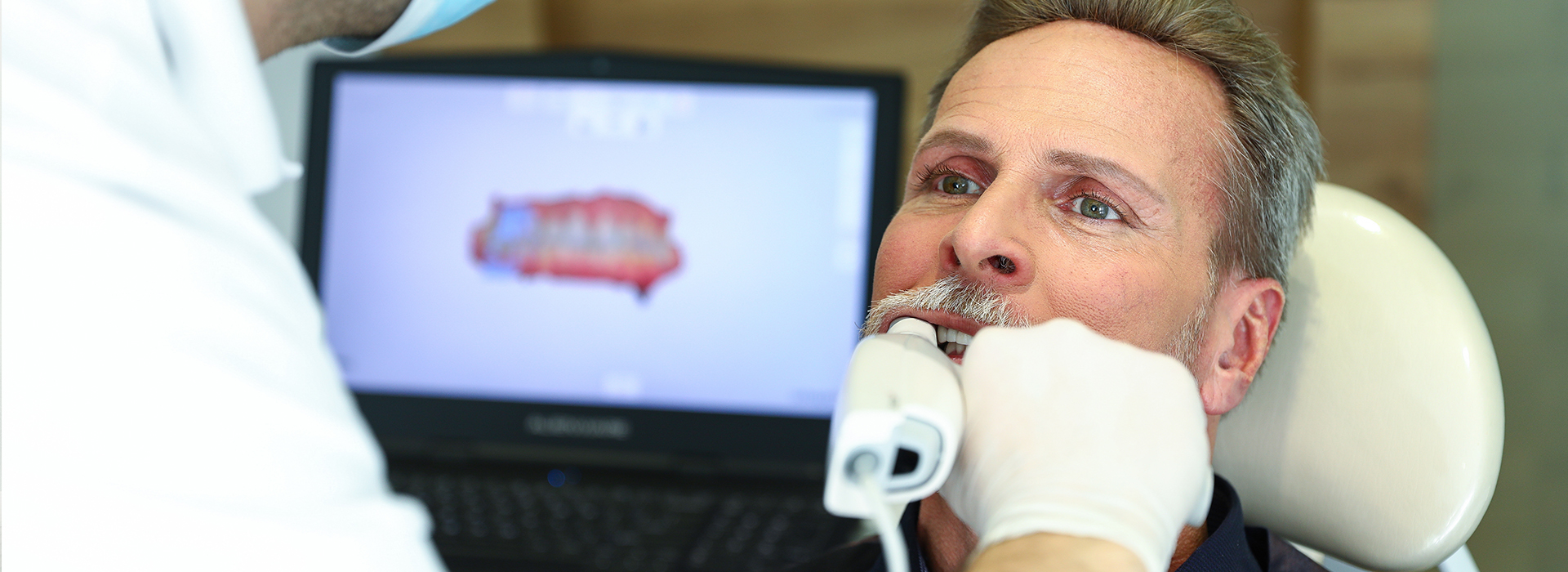
Digital impressions use small, handheld intraoral scanners to capture a detailed 3D image of teeth and surrounding tissues. Instead of bite trays and impression putty, the clinician gently moves the scanner around the mouth while software stitches hundreds of optical images into a single, accurate model. The result is an immediate, computer-generated representation that can be reviewed on-screen in real time.
For patients, the experience is noticeably different: scans are quick, dry, and free from the gag-inducing feel of conventional materials. For clinicians, the immediate visual feedback allows for confirmation of margins, contacts, and soft-tissue relationships before the patient leaves the chair. This combination of comfort and speed is why many practices are transitioning from analog molds to digital workflows.
Beyond the simple convenience, digital impressions form the foundation for many contemporary restorative and orthodontic processes. Their flexibility supports everything from single crowns and veneers to multiunit prostheses and appliance fabrication, making them a versatile tool in modern dental care.
One of the clearest benefits patients report is the absence of impression material. No more bulky trays, unpleasant flavors, or extended periods of stillness while materials set. This is particularly helpful for those with strong gag reflexes or limited tolerance for long, uncomfortable procedures.
Digital scanning also improves communication during the visit. Because scans appear instantly on a monitor, clinicians can show patients exactly what they see—areas of wear, margins that need attention, or spaces needing restoration. This visual approach helps patients understand treatment options and fosters shared decision-making.
From a practical standpoint, scans often shorten appointment time. Many steps associated with traditional impressions—mixing materials, waiting for sets, and preparing stone models—are eliminated. The patient experiences a smoother visit, and the care team gains time to focus on precise treatment planning and patient education.
Accuracy is a primary reason dental teams adopt digital impressions. High-resolution scanners capture fine anatomic details and occlusal relationships that are critical for well-fitting crowns, bridges, and onlays. When the digital model is accurate, technicians and CAD/CAM systems can fabricate restorations that require fewer adjustments at try-in.
Digital files also reduce the risk of distortions and inaccuracies that can occur with traditional impression materials and stone models. Since the scan is a direct optical capture, it is not subject to dimensional changes from setting expansion or shrinkage—common sources of fit problems in analog workflows.
For complex restorative cases, precise digital impressions make it easier to plan margin designs, evaluate interproximal contacts, and check clearances for opposing dentition. This level of detail contributes to smoother restorative appointments and outcomes that align more closely with the treatment plan.
Once a scan is complete, the data can be used in several ways depending on the treatment goal. Files can be sent electronically to an external dental laboratory, imported into in-office CAD software for design, or combined with cone-beam CT images for implant planning. Electronic transmission accelerates collaboration and removes delays associated with shipping physical impressions.
In practices equipped with in-office milling or 3D printing, scans can lead directly to same-day restorations. The clinician or technician designs the restoration using CAD software, fabricates it chairside, and delivers it in a single visit when appropriate. Even when a lab completes the work, the streamlined digital handoff reduces turnaround time and minimizes the chance for errors in communication.
Digital workflows also facilitate better recordkeeping. Electronic models are stored without physical space constraints and can be revisited for future restorative needs or comparative assessments. This makes follow-up care and long-term planning more efficient and data-driven.
Additionally, the use of standardized digital formats improves interoperability between different labs and systems. Most modern labs accept common file types, which simplifies the transfer of information and helps ensure the scanned data is interpreted consistently.
Digital impressions work well for many restorative and prosthetic indications, including single crowns, inlays/onlays, veneers, implant restorations, and orthodontic records. They are particularly advantageous when precise margin visualization and occlusal relationships are essential to the outcome.
There are clinical situations that require careful technique: heavy bleeding, excessive saliva, or limited access can challenge optical capture and may necessitate additional isolation or preparation. Experienced clinicians know how to manage these variables—using gentle retraction, moisture control, and incremental scanning—to produce reliable results.
Some restorative decisions also factor into the choice of scanning versus traditional impressions. In complex full-arch reconstructions or cases requiring certain material workflows, the dental team will evaluate the best approach and may combine digital techniques with other methods when indicated. The goal is always a predictable, high-quality result that aligns with the patient’s needs.
At Murphy Dentistry, our team integrates digital impressions into patient care to enhance comfort, accuracy, and efficiency. If you’d like to learn more about how digital scanning may help streamline your restorative or cosmetic treatment, please contact us for more information.

Taking the next step toward your ideal smile is simple. Whether you're ready to schedule your appointment or simply have questions about our services or treatment options, our friendly staff is here to help.