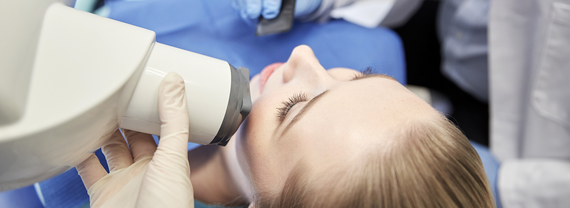
Digital radiography is the contemporary approach to dental imaging that replaces traditional film with electronic sensors and computer processing. Instead of exposing a physical film to X-rays and developing it in a darkroom, a digital sensor captures the image instantly and converts it into a digital file. This shift has redefined how clinicians evaluate dental health, allowing clearer visualization and more flexible manipulation of images without the delays associated with film.
For patients, the change is more than technological—it alters the pace and clarity of care. Where film required handling time and chemical processing, digital images appear on a monitor within seconds. That immediacy supports faster diagnosis and supports collaborative conversations between patient and provider about findings and treatment options.
Adoption of digital radiography is now a standard in many progressive practices because it streamlines clinical workflows and enables more precise record keeping. In addition to improving day-to-day efficiency, the digital format facilitates secure storage and straightforward sharing with specialists or other providers when comprehensive care is needed.
At the heart of digital radiography are flat-panel detectors or charge-coupled devices (CCDs) that act as direct substitutes for film. These sensors detect X-ray photons and convert them into digital signals, which software then renders as high-resolution images. Unlike film, the output can be adjusted for contrast, brightness, and magnification, helping clinicians identify subtle changes in enamel, dentin, and bone.
Software tools allow clinicians to zoom in, measure distances, and apply filters that enhance specific features of the image without re-exposing the patient. This capability is especially useful for detecting early-stage decay, evaluating root canal anatomy, and monitoring periodontal bone levels. Because clinicians can fine-tune the image display, fewer retakes are typically necessary, preserving both diagnostic value and patient comfort.
Digital images can be annotated and integrated into electronic health records, so all image-based findings become part of the comprehensive patient chart. This continuity supports ongoing care by providing a clear, time-stamped visual history that clinicians can consult when planning restorative, surgical, or preventative treatments.
One of the most tangible benefits of digital radiography is the reduction in radiation exposure compared with conventional film-based X-rays. Digital sensors are more sensitive to X-rays than film, which means high-quality images can be produced with lower doses. Regulatory bodies and professional organizations encourage the use of techniques and technologies that minimize exposure while maintaining diagnostic utility, and digital systems align with that goal.
Lower radiation per image is particularly relevant for patients who require frequent monitoring, such as those undergoing orthodontic treatment, periodontal maintenance, or follow-up after implant placement. By reducing cumulative exposure without sacrificing diagnostic detail, digital radiography supports long-term patient safety while enabling necessary clinical oversight.
Additionally, digital workflows eliminate chemical processing and its associated waste, making digital radiography a more environmentally responsible choice. The absence of film developers and fixers reduces hazardous waste, aligning imaging practices with broader goals of sustainability in healthcare settings.
Digital radiography accelerates the clinical workflow by producing instant images that can be reviewed chairside. This immediacy shortens appointments that depend on radiographic assessment and enables real-time discussion of findings with patients. Clinicians can show images on a monitor, point out concerns, and review proposed treatment steps with clear visual context, which improves patient understanding and informed decision-making.
From a practice management perspective, digital images are easy to archive, retrieve, and share. When a patient needs referral care, images can be transmitted electronically to a specialist or another office, facilitating timely consultations and coordinated treatment planning. This capability reduces delays that once occurred when physical films had to be mailed or hand-delivered.
Digital archiving also simplifies long-term monitoring. Clinicians can compare current images with historical ones side by side, track subtle changes over time, and document the progression of conditions such as caries, bone loss, or healing around implants. The result is a more cohesive record that supports continuity of care across multiple visits and providers.
Digital radiography is often used alongside other modern imaging modalities—such as intraoral scanners and cone-beam computed tomography (CBCT)—to provide a full picture of oral health. While 2D digital X-rays excel at many routine diagnostic tasks, three-dimensional imaging adds critical detail for complex implant planning, surgical evaluations, and assessment of anatomical structures. Together, these tools enable more predictable, evidence-based treatment outcomes.
Clinicians trained in digital imaging can leverage these tools to make more confident diagnoses and design treatment plans that reflect the patient’s specific anatomy. The interoperability of digital files means that results from radiographs, scans, and intraoral images can be combined to form a richer clinical narrative than any single image could provide.
At Murphy Dentistry, we integrate digital radiography into our diagnostic and treatment workflows to support precise care across preventive, restorative, and surgical services. The technology helps ensure that recommendations are grounded in clear visual evidence, and it supports efficient coordination when multiple specialists are involved in a patient’s care.
In summary, digital radiography represents a meaningful advancement in dental imaging: it enhances diagnostic clarity, reduces radiation exposure, streamlines clinical workflows, and dovetails with other modern diagnostic tools. If you’d like to learn more about how digital imaging is used in dental care or how it might apply to your own treatment, please contact us for more information.

Taking the next step toward your ideal smile is simple. Whether you're ready to schedule your appointment or simply have questions about our services or treatment options, our friendly staff is here to help.